The Nervous System & Sense Organs

ICSE Class 10 Biology · Goyal New Approach Biology · 95 solved questions

Exercise
1Test Yourself 1

Name the following:

(i) Neurons convey impulse from receptor to the brain.

(ii) Neurons connecting sensory and motor neuron.

(iii) Inflammation of the meninges.

(iv) Matter present in the inner portion of cerebrum.

(v) A band of nerve fibre present between two cerebral hemispheres.

Show SolutionHide Solution↓

(i) Sensory (afferent) neurons

(ii) Connector (association) neurons

(iii) Meningitis

(iv) White matter

(v) Corpus callosum

2Test Yourself 1

Mention if the following statements are True/False:

(i) Spinal cord conducts both sensory as well as motor responses.

(ii) Olfactory nerve is a sensory nerve.

(iii) Grey matter of cerebrum is composed of axons of the neurons.

(iv) Medulla oblongata maintains equilibrium.

(v) Peristaltic reflexes are the examples of simple reflexes.

Show SolutionHide Solution↓

(i) True

(ii) True

(iii) False

(iv) False

(v) True

1Test Yourself 2

Fill in the blanks:

(i) Yellow spot of eye is also known as ............... .

(ii) No image is formed in ............... .

(iii) ............... are photoreceptor cells.

(iv) Pigment present in cone cell is ............... .

(v) Inner fibrous coat of eye ball is ............... .

Show SolutionHide Solution↓

(i) Yellow spot of eye is also known as macula or fovea centralis.

(ii) No image is formed in blind spot.

(iii) Rods and cones are photoreceptor cells.

(iv) Pigment present in cone cell is iodopsin.

(v) Inner fibrous coat of eye ball is retina.

2Test Yourself 2

Mention if the following statements are true (T) or false (F):

(i) Ciliary muscles change the thickness of the lens when focusing.

(ii) The conjunctiva covers the entire front part of the eye.

(iii) Rods are responsible for colour vision.

(iv) Blind spot is also called fovea centralis.

(v) Astigmatism is corrected by using biconvex lens.

Show SolutionHide Solution↓

(i) True

(ii) True

(iii) False

(iv) False

(v) False

1Test Yourself 3

Write the functions of following:

(i) Ear ossicle

(ii) Semicircular canals

(iii) Cochlea

(iv) Eustachian tube

Show SolutionHide Solution↓

(i) Ear ossicle — The three ear ossicles (malleus, incus and stapes) transmit the vibrations from the ear drum to the oval window and amplify them due to their lever-like action.

(ii) Semicircular canals — They help in maintaining the dynamic equilibrium (balance during movement) of the body.

(iii) Cochlea — It contains the organ of Corti, which converts sound vibrations into nerve impulses that are transmitted to the brain via the auditory nerve, thus helping in hearing.

(iv) Eustachian tube — It equalises the air pressure on either side of the ear drum, allowing it to vibrate freely.

2Test Yourself 3

Write in proper sequence, the names of all the parts of the human ear through which sound waves move.

Show SolutionHide Solution↓

Pinna → auditory canal → ear drum (tympanum) → ear ossicles (malleus, incus, stapes) → oval window → cochlea → auditory nerve

1Choose Most Appropriate Answer

Which of the following is not a natural reflex action?

Show SolutionHide Solution↓

Salivation at the sight of food

Reason — Salivation at the sight of food is a conditioned (acquired) reflex, learnt during the lifetime, and not an inborn natural reflex. Knee-jerk, blinking due to strong light and sneezing are all inborn, unconditioned (natural) reflexes.

2Choose Most Appropriate Answer

The ventral root ganglion of the spinal cord contains cell bodies of the

Show SolutionHide Solution↓

Motor neuron

Reason — The ventral root contains motor (efferent) fibres that arise from motor neurons whose cell bodies are located in the ventral horn of the grey matter of the spinal cord.

3Choose Most Appropriate Answer

The two cerebral hemispheres are joined by a sheet of fibres called:

Show SolutionHide Solution↓

Corpus callosum

Reason — The two cerebral hemispheres are connected together by a sheet of nerve fibres called corpus callosum.

4Choose Most Appropriate Answer

Assertion (A): The point of contact between two neurons is called synapse.

Reason (R): Synapse is formed between the dendrites of adjacent neurons.

Show SolutionHide Solution↓

(A) is true and (R) is false.

Explanation The synapse is the point of contact between the terminal branches of the axon of one neuron and the dendrites of another neuron. It is formed between the axon endings of one neuron and the dendrites of the next neuron, not between the dendrites of adjacent neurons.

5Choose Most Appropriate Answer

Assertion (A): Rods and Cones are photoreceptors in the sclera of eyeball.

Reason (R): Rods are sensitive to dim light.

Show SolutionHide Solution↓

(A) is false and (R) is true.

Explanation Rods and cones are photoreceptor cells present in the retina, not in the sclera. Rods are indeed sensitive to dim light and are important for night vision.

6Choose Most Appropriate Answer

Which one of the following is mainly associated with the maintenance of the posture?

Show SolutionHide Solution↓

Cerebellum

Reason — The cerebellum coordinates voluntary muscular movements and maintains posture, equilibrium and muscular toning of the body.

7Choose Most Appropriate Answer

The part of eye which is grafted in a needy patient from a donated eye, is

Show SolutionHide Solution↓

Cornea

Reason — The cornea is a transparent, non-vascular part in front of the eye. It can be donated from a clinically dead person and grafted in a needy patient to restore vision.

8Choose Most Appropriate Answer

Assertion (A): Arachnoid layer helps in speeding up the transmission of nerve impulses along the axons.

Reason (R): Axon endings at the synapse secrete acetylene.

Show SolutionHide Solution↓

Both (A) and (R) are false.

Explanation The arachnoid is the middle layer of the meninges that protects the brain and spinal cord; it does not speed up the transmission of nerve impulses. Speed of impulse conduction is increased by the myelin sheath. Also, the axon endings at the synapse secrete a neurotransmitter such as acetylcholine, not acetylene.

9Choose Most Appropriate Answer

Stimulation of the nerves of the sympathetic nervous system

Show SolutionHide Solution↓

Accelerates heartbeat

Reason — The sympathetic nervous system prepares the body for emergency action, so it accelerates the heartbeat. The remaining options (constriction of pupil, increased peristalsis and retarding of heartbeat) are actions of the parasympathetic nervous system.

10Choose Most Appropriate Answer

Lata wanted to cross the road. She looked on either side of the road and then walked across to the other side of the road.

Which of the following is/are involved in the process described above?

i. Cerebrum ii. Cerebellum iii. Skeletal muscles iv. Medulla Oblongata

Show SolutionHide Solution↓

Only i, ii and iii

Reason — Looking and deciding to cross is a voluntary action controlled by the cerebrum, walking involves the skeletal muscles, and the cerebellum coordinates the muscular movements and balance during walking.

11Choose Most Appropriate Answer

The membranes covering the brain are called

Show SolutionHide Solution↓

meninges

Reason — The brain and spinal cord are covered by three protective membranes collectively called meninges (dura mater, arachnoid and pia mater).

12Choose Most Appropriate Answer

The number of spinal nerves in a human being are

Show SolutionHide Solution↓

31 pairs

Reason — A human body has 31 pairs of spinal nerves.

13Choose Most Appropriate Answer

The vibrations received by outer ear are transported through the auditory canal to

Show SolutionHide Solution↓

eardrum

Reason — The pinna collects the sound waves and directs them through the auditory canal, which terminates at the ear drum, causing it to vibrate.

14Choose Most Appropriate Answer

A defect in which parallel rays from the infinity are focussed behind the retina is called

Show SolutionHide Solution↓

hypermetropia

Reason — In hypermetropia (long-sightedness), the image is formed behind the retina, so nearby objects appear blurred while distant vision is clear.

15Choose Most Appropriate Answer

Which one of these looks like a snail's shell?

Show SolutionHide Solution↓

cochlea

Reason — The cochlea is a fluid-filled, spiral-shaped tube resembling a snail's shell.

16Choose Most Appropriate Answer

Rahul inserted a hairpin into his right ear to remove ear wax. He felt a sudden sharp pain with loss of hearing. This was due to:

Show SolutionHide Solution↓

Rupture of eardrum

Reason — Inserting a sharp object into the ear can rupture the thin ear drum, causing sudden sharp pain and loss of hearing.

17Choose Most Appropriate Answer

While playing with his friends, Peter inserted a stick into his ear. He lost his hearing due to the rupture of:

Show SolutionHide Solution↓

Ear drum

Reason — The ear drum is a thin membrane that vibrates on receiving sound waves. Inserting a stick can rupture it, leading to loss of hearing.

18Choose Most Appropriate Answer

The structure that lies at the junction of the choroid and the iris and is itself a part of the choroid is called:

Show SolutionHide Solution↓

Ciliary body

Reason — The ciliary body is the thickest portion of the choroid, arising from a point just behind the junction of the sclera and the cornea, and lies at the region of the iris.

19Choose Most Appropriate Answer

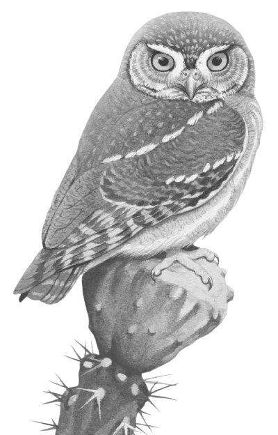
Owls are nocturnal animals. They are active at night and hunt small animals for food. Owls have large eyes and can see well at night. This is due to:

Owls are nocturnal animals. They are active at night and hunt small animals for food. Owls have large eyes and can see well at night. This is due to: ICSE 2025 Biology Solved Question Paper.
Show SolutionHide Solution↓

Rods and Rhodopsin

Reason — Owls see well at night because their eyes have many rods, which work in dim light. Rods contain rhodopsin, a pigment that helps detect light in the dark.

20Choose Most Appropriate Answer

Spinal cord originates from

Show SolutionHide Solution↓

Medulla

Reason — The spinal cord extends from the medulla oblongata to the lumbar region through the neural canal of the vertebral column.

21Choose Most Appropriate Answer

Vitreous humour is present between

Show SolutionHide Solution↓

Lens and Retina

Reason — The vitreous chamber lies behind the lens and is filled with the jelly-like vitreous humour, occupying the space between the lens and the retina.

22Choose Most Appropriate Answer

The number of cranial nerves in humans are

Show SolutionHide Solution↓

12 pairs

Reason — A human body has 12 pairs of cranial nerves.

23Choose Most Appropriate Answer

The circular opening in the centre of iris

Show SolutionHide Solution↓

Pupil

Reason — The pupil is the circular window (opening) lying in the centre of the iris, through which light enters the eye.

24Choose Most Appropriate Answer

Organ of corti is present inside the

Show SolutionHide Solution↓

Cochlea

Reason — The middle canal of the cochlea contains the sound reception system called the organ of Corti.

25Choose Most Appropriate Answer

Aqueous humour is present between the:

Show SolutionHide Solution↓

Cornea and Lens

Reason — The aqueous chamber is present between the lens and the cornea and is filled with the watery fluid called aqueous humour.

1Very Short Answer Questions

Name the following:

(i) The part of the brain that regulates the peristaltic movement of the alimentary canal.

(ii) The collective term for the protective membranes of the brain.

(iii) A structure that collects the sound waves and conducts them through the external auditory canal.

(iv) Three tiny bones present in the middle ear.

(v) The fibres which hold the lens of the eye in position.

(vi) The glands located at the upper sideward portion of the orbit of eye.

(vii) The layer of the eyeball that provides nourishment to the eye.

(viii) The lens of eye losing flexibility resulting in a kind of longsightedness in middle aged people.

(ix) The part of the brain that carries impulses from one hemisphere of the cerebellum to the other.

(x) Maintaining balance of the body and coordinating muscular activities is carried out by.

(xi) The part of the eye which can be donated from a clinically dead person.

(xii) A tubular passage that connects the pharynx and the middle ear.

Show SolutionHide Solution↓

(i) Medulla oblongata

(ii) Meninges

(iii) Pinna

(iv) Ear ossicles (malleus, incus and stapes)

(v) Suspensory ligaments

(vi) Lacrimal glands

(vii) Choroid

(viii) Presbyopia

(ix) Pons

(x) Cerebellum

(xi) Cornea

(xii) Eustachian tube

2Very Short Answer Questions

Fill in the blanks with suitable words:

The technical term for short sightedness is (a)............... This defect is caused because the eyeball is (b)............... from front to back or the lens is too (c)............... . It can be corrected by using a suitable (d)............... lens. The power of the lens is mentioned in (e)............... .

Show SolutionHide Solution↓

The technical term for short sightedness is (a) myopia. This defect is caused because the eyeball is (b) lengthened from front to back or the lens is too (c) convex. It can be corrected by using a suitable (d) concave lens. The power of the lens is mentioned in (e) minus (–).

3Very Short Answer Questions

Choose the appropriate answer.

(i) Which one surrounds the organ of Corti? Perilymph or endolymph.

(ii) Which one forms the iris? Sclerotic layer or Choroid layer.

Show SolutionHide Solution↓

(i) Endolymph

(ii) Choroid layer

4Very Short Answer Questions

Mention if the following statements are True/False. If false, rewrite the wrong ones in the correct form by changing only the words printed in bold face:

(i) Cones enable us to see three primary colours.

(ii) Cranial nerves arise from the brain.

(iii) Rods are responsible for vision in the dark.

(iv) All voluntary actions are controlled by the cerebellum.

(v) Dilation of the pupil is brought about by the sympathetic nervous system.

(vi) The outermost layer of meninges is pia mater.

Show SolutionHide Solution↓

(i) True

(ii) True

(iii) True

(iv) False
Corrected Statement — All voluntary actions are controlled by the cerebrum.

(v) True

(vi) False
Corrected Statement — The outermost layer of meninges is dura mater.

5Very Short Answer Questions

Arrange and rewrite the terms in each group in correct order to be in a logical sequence, beginning with the term that is underlined.

(i) Receptor, Response, Effector, Spinal cord

(ii) Fovea, Lens, Cornea, Conjunctiva

(iii) Cochlea, Malleus, Pinna, Stapes

(iv) Auditory canal, Organ of Corti, Stapes, Malleus.

Show SolutionHide Solution↓

(i) Receptor → Spinal cord → Effector → Response

(ii) Conjunctiva → Cornea → Lens → Fovea

(iii) Pinna → Malleus → Stapes → Cochlea

(iv) Auditory canal → Malleus → Stapes → Organ of Corti

6Very Short Answer Questions

Choose the odd one out from the following terms and name the category to which the others belong.

(i) Oval window, Cochlea, Auditory canal, Round window

Show SolutionHide Solution↓

(i) Odd term: Auditory canal
Category: Parts of the inner ear

7Very Short Answer Questions

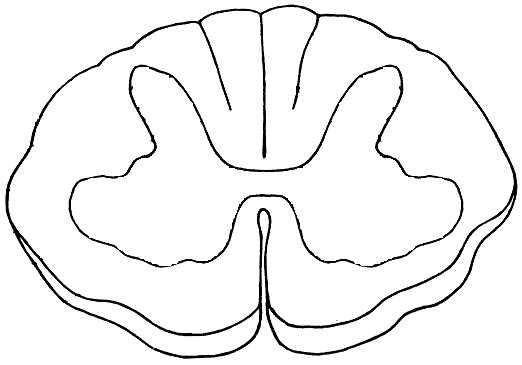
Given below is the transverse section of the spinal cord. Read the information below the diagram and fill in the blanks:

Given below is the transverse section of the spinal cord. Read the information below the diagram and fill in the blanks: ICSE 2025 Biology Solved Question Paper.

The spinal cord extends from the medulla oblongata of the brain and runs down through the whole length of the vertebral column. The spinal cord is covered by the meninges. It conducts impulses from the skin and muscles to the brain. It also conducts impulses from the brain to the muscles of the trunk and limbs.

The spinal cord is a part of the (a) ............... Nervous System. The grey matter in the picture given above consists of (b) ............... while the white matter consists of (c) ............... . The spinal cord is concerned with the (d) ............... actions below the neck. (e) ............... is the bony structure that protects the spinal cord.

Show SolutionHide Solution↓

The spinal cord is a part of the (a) Central Nervous System. The grey matter in the picture given above consists of (b) cytons while the white matter consists of (c) axons. The spinal cord is concerned with the (d) reflex actions below the neck. (e) Vertebral column is the bony structure that protects the spinal cord.

8Very Short Answer Questions

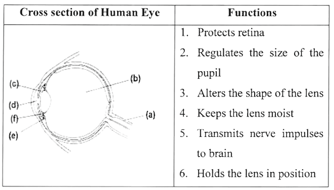
Given below is a cross section of the human eye. Match the structures marked (a) to (e) with their correct functions:

Example: (f) - 6. Holds the lens in position

Given below is a cross section of the human eye. ICSE 2025 Biology Solved Question Paper.
Show SolutionHide Solution↓

(a) - 5. Transmits nerve impulses to brain

(b) - 1. Protects retina

(c) - 2. Regulates the size of the pupil

(d) - 4. Keeps the lens moist

(e) - 3. Alters the shape of the lens

1(i)Short Answer Questions

Define Reflex action.

Show SolutionHide Solution↓

Reflex action is a rapid, automatic, involuntary response to a stimulus, for example, immediate withdrawal of the hand on touching a hot object.

1(ii)Short Answer Questions

Define Power of accommodation.

Show SolutionHide Solution↓

Power of accommodation is the ability of the eye to adjust the curvature (and hence focal length) of the elastic lens so as to obtain a clear vision of objects at varying distances.

1(iii)Short Answer Questions

Define Vitreous humour.

Show SolutionHide Solution↓

Vitreous humour is the transparent, jelly-like substance (containing water and salt) filling the vitreous chamber behind the lens. It helps to maintain the shape of the eyeball and protects the retina.

1(iv)Short Answer Questions

Define Stereoscopic vision.

Show SolutionHide Solution↓

Stereoscopic (binocular) vision is the ability to perceive depth or the relative distance of an object due to the simultaneous focusing of the object by both eyes, whose two images are correlated by the brain into a single three-dimensional impression.

1(v)Short Answer Questions

Define Synapse.

Show SolutionHide Solution↓

Synapse is the junctional region between two neurons where information from one neuron is transmitted (handed over) to another neuron, although there is no protoplasmic connection between the two neurons.

1(vi)Short Answer Questions

Define Lacrimal gland.

Show SolutionHide Solution↓

Lacrimal gland is the gland located at the upper, outer end of the eyeball, beneath the eyelid, which secretes tears that serve as a lubricant, wash away dust particles and have antiseptic property due to the enzyme lysozyme.

2(i)Short Answer Questions

Differentiate between Rods and cones. (Pigment present)

Show SolutionHide Solution↓

Differences between Rods and cones are:

RodsCones
Rods contain a pigment called rhodopsin.Cones contain a pigment called iodopsin.
2(ii)Short Answer Questions

Differentiate between Semicircular canals and utriculus. (Function)

Show SolutionHide Solution↓

Differences between Semicircular canals and utriculus are:

Semicircular canalsUtriculus
Semicircular canals are concerned with dynamic equilibrium (balance during movement).Utriculus is concerned with static equilibrium (balance in response to gravity).
2(iii)Short Answer Questions

Differentiate between Dynamic balance and static balance. (Definition)

Show SolutionHide Solution↓

Differences between Dynamic balance and static balance are:

Dynamic balanceStatic balance
It is the maintenance of body position in response to movement (motion).It is the maintenance of body position in response to gravity (at rest).
2(iv)Short Answer Questions

Differentiate between Myopia and hypermetropia. (condition of eyeball)

Show SolutionHide Solution↓

Differences between Myopia and hypermetropia are:

MyopiaHypermetropia
It is caused by elongation (lengthening) of the eyeball from front to back or the lens becoming too convex.It is caused by shortening of the eyeball from front to back or the lens becoming too flat.
2(v)Short Answer Questions

Differentiate between Spinal nerves and cranial nerves. (Number of nerves)

Show SolutionHide Solution↓

Differences between Spinal nerves and cranial nerves are:

Spinal nervesCranial nerves
There are 31 pairs of spinal nerves.There are 12 pairs of cranial nerves.
2(vi)Short Answer Questions

Differentiate between Near vision and distant vision. (Shape of the eye lens)

Show SolutionHide Solution↓

Differences between Near vision and distant vision are:

Near visionDistant vision
For near vision, the lens becomes more rounded (convex).For distant vision, the lens becomes flat (thin).
2(vii)Short Answer Questions

Differentiate between Semicircular and cochlea. (Sense)

Show SolutionHide Solution↓

Differences between Semicircular canals and cochlea are:

Semicircular canalsCochlea
Semicircular canals are concerned with the sense of balance (equilibrium).Cochlea is concerned with the sense of hearing.
3Short Answer Questions

Mention the location and function of the following:

(i) Cerebellum

(ii) Cerebrospinal fluid

(iii) Pinna

(iv) Iris of the eye

(v) Eustachian tube

(vi) Myelin Sheath

(vii) Suspensory ligaments of the eye

(viii) Cerebrum

(ix) Lacrimal gland

(x) Malleus

(xi) Incus

(xii) Corpus callosum

(xiii) Medulla oblongata

(xiv) Dura mater

Show SolutionHide Solution↓

(i) Cerebellum

Location — It lies below the cerebrum and above the medulla, in the hindbrain.

Function — It maintains posture, equilibrium and muscular toning, and coordinates voluntary muscular movements.

(ii) Cerebrospinal fluid

Location — It is present between the meninges (in the space between the inner two layers), in the ventricles of the brain and in the central canal of the spinal cord.

Function — It acts as a shock-absorbing cushion, protects the brain and spinal cord from injury, supplies nutrients and removes wastes.

(iii) Pinna

Location — It is the fleshy, cup-shaped part of the outer ear.

Function — It collects and directs sound waves towards the auditory canal (ear drum).

(iv) Iris

Location — It is the coloured part of the choroid, present around the pupil in front of the eye.

Function — By the contraction of its radial and circular muscles, it regulates the size of the pupil and thus controls the amount of light entering the eye.

(v) Eustachian tube

Location — It connects the cavity of the middle ear with the pharynx (throat).

Function — It equalises the air pressure on either side of the ear drum, allowing it to vibrate freely.

(vi) Myelin sheath

Location — It is the white, multi-layered covering around the axon of a nerve fibre.

Function — It increases the speed of nerve impulse conduction and insulates the axon, preventing mixing of impulses in the adjacent fibres.

(vii) Suspensory ligaments

Location — They are present between the lens and the ciliary body.

Function — They hold the lens in position and attach it to the ciliary body.

(viii) Cerebrum

Location — It is the largest, uppermost part of the forebrain.

Function — It is the seat of consciousness, thinking, memory, reasoning, intelligence and perception, and controls voluntary actions.

(ix) Lacrimal gland

Location — It is located at the upper, outer end of the eyeball, beneath the eyelid.

Function — It secretes tears that lubricate the eye, wash away dust particles and have antiseptic property.

(x) Malleus

Location — It is the first (hammer-shaped) ear ossicle of the middle ear, attached to the inner surface of the ear drum.

Function — It transmits and amplifies the vibrations of the ear drum to the incus.

(xi) Incus

Location — It is the second (anvil-shaped) ear ossicle of the middle ear, present between the malleus and the stapes.

Function — It transmits and amplifies the vibrations from the malleus to the stapes.

(xii) Corpus callosum

Location — It is a sheet of nerve fibres present in the forebrain, connecting the two cerebral hemispheres.

Function — It connects the two cerebral hemispheres and transfers information from one hemisphere to the other.

(xiii) Medulla oblongata

Location — It is the lowest part of the brain, located at the base of the skull, continuing below with the spinal cord.

Function — It contains centres controlling cardiac, respiratory and vasomotor activities, and reflex centres for swallowing, coughing, sneezing and vomiting.

(xiv) Dura mater

Location — It is the outermost of the three meninges covering the brain and spinal cord.

Function — It gives tough protective covering to the brain and spinal cord.
4(i)Short Answer Questions

Give reason — The hand automatically shows the direction to turn a cycle without thinking.

Show SolutionHide Solution↓

Riding a cycle and turning it in the correct direction is a conditioned reflex, which is acquired through repeated practice during the lifetime. Once learnt, the action becomes automatic and is performed without conscious thinking.

4(ii)Short Answer Questions

Give reason — Injury to the medulla oblongata results in death.

Show SolutionHide Solution↓

The medulla oblongata contains the vital reflex centres that control cardiac (heartbeat), respiratory (breathing) and vasomotor activities. Any injury to it stops these essential involuntary functions, which results in death.

4(iii)Short Answer Questions

Give reason — A person after consuming alcohol walks clumsily.

Show SolutionHide Solution↓

Alcohol affects the cerebellum, which is responsible for coordinating muscular movements and maintaining the balance of the body. As a result, the person cannot coordinate the movements of the head, eyes and limbs properly and hence walks clumsily.

4(iv)Short Answer Questions

Give reason — One feels blinded for a short time while coming out of a dark room.

Show SolutionHide Solution↓

In the dark, the pupil is dilated and the pigment rhodopsin is regenerated in the rods. On coming into bright light suddenly, the excess rhodopsin gets rapidly broken down and the pupil takes some time to constrict. During this short adjustment (light adaptation), the person feels blinded for a short while.

4(v)Short Answer Questions

Give reason — We cannot distinguish colours in moonlight.

Show SolutionHide Solution↓

Colour vision is provided by the cone cells, which are sensitive only to bright light. In dim moonlight, the cones do not function; only the rods (sensitive to dim light) work, and rods do not respond to colour. Hence, we cannot distinguish colours in moonlight.

5(i)Short Answer Questions

Which is the light sensitive layer of the eyeball? Where exactly is the image formed on this layer?

Show SolutionHide Solution↓

The retina is the light-sensitive layer of the eyeball. The image is formed on the retina and is most distinct at the yellow spot (fovea centralis), the region of best vision.

5(ii)Short Answer Questions

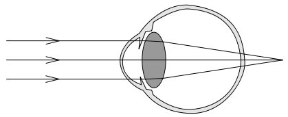
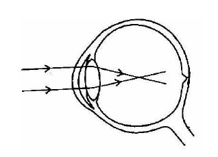
Study the diagram given below and answer the questions that follow :

Identify the defect of the eye by mentioning the technical term. Mention one reason for this defect. Name the type of lens used to correct this defect. ICSE 2022 Biology Solved Question Paper.

(a) Identify the defect of the eye by mentioning the technical term.

(b) Mention one reason for this defect.

(c) Name the type of lens used to correct this defect.

Show SolutionHide Solution↓

(a) Hyperopia.

(b) Lens is too flat.

(c) Convex lens.

5(iii)Short Answer Questions

How are cytons and axons of neurons placed in the cerebrum?

Show SolutionHide Solution↓

In the cerebrum, the outer region (cortex) contains the cell bodies (cytons) of the neurons and is therefore the grey matter, while the inner region contains the axons of the neurons and forms the white matter.

5(iv)Short Answer Questions

Name the nerve that transmits impulses to the brain from :

(a) Ear

(b) Eye

Show SolutionHide Solution↓

(a) Auditory nerve

(b) Optic nerve

5(v)Short Answer Questions

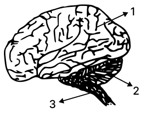
The diagram given below is that of a human brain. Answer the questions that follow :

The diagram shows a human brain. Label the parts numbered 2 and 3. State any one function of the part numbered 1. ICSE 2022 Biology Solved Question Paper.

(a) Label the parts numbered 2 and 3.

(b) State any one function of the part numbered 1.

Show SolutionHide Solution↓

(a) Parts numbered 2 and 3 are:

  • 2 → Cerebellum
  • 3 → Medulla oblongata

(b) The part labelled as 1 is Cerebrum. It is the seat of intelligence, consciousness and will-power. It controls all voluntary actions.

5(vi)Short Answer Questions

What is a reflex action? Name the two types of reflexes.

Show SolutionHide Solution↓

Reflex action is an automatic/quick/immediate, involuntary action in the body brought about by a stimulus. The two types of reflexes are natural reflexes and conditioned reflexes.

5(vii)Short Answer Questions

Write the two functions of cerebellum.

Show SolutionHide Solution↓

The two functions of cerebellum are:

  1. Maintains balance of the body.
  2. Coordinates muscular activity.
5(viii)Short Answer Questions

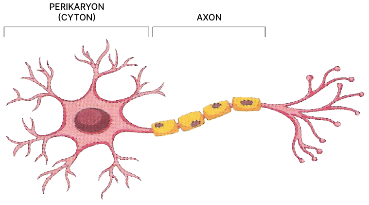
Draw a neat diagram of a Neuron and label any two parts.

Show SolutionHide Solution↓

Diagram of Neuron with Cyton and Axon labelled is shown below:

Draw a neat diagram of a Neuron and label any two parts. Nervous System, ICSE Class 10 Goyal Biology Solutions.
5(ix)Short Answer Questions

Name the two sensory cells in retina meant for light adaptation.

Show SolutionHide Solution↓

Rod cells and cone cells are responsible for light adaptations.

6Short Answer Questions

Rachna is an expert gymnast on the balance beam. She has been vigorously practising for the Olympic Games. Lately, she has been experiencing dizziness and losing her balance during the practice sessions.

Rachna is an expert gymnast on the balance beam. She has been vigorously practising for the Olympic Games. Lately, she has been experiencing dizziness and losing her balance during the practice sessions. ICSE 2025 Biology Solved Question Paper.

(a) Which part of the membranous labyrinth is responsible for maintaining the body balance on the beam?

(b) Which type of balance is maintained by the structure mentioned in (a)?

Show SolutionHide Solution↓

(a) The semicircular canals.

(b) Dynamic balance.

7Short Answer Questions

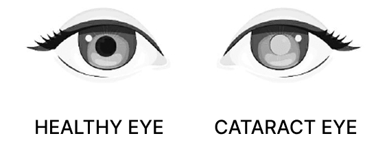
Mr. Sharma is a 76 year old man. His vision was gradually becoming blurred. He consulted an eye doctor and it was diagnosed as cataract.

Mr. Sharma is a 76 year old man. His vision was gradually becoming blurred. He consulted an eye doctor and it was diagnosed as cataract. ICSE 2025 Biology Solved Question Paper.

(a) Which part of the eye does cataract affect?

(b) Why does the vision become blurred?

Show SolutionHide Solution↓

(a) Lens

(b) The vision becomes blurred because the lens becomes cloudy or opaque, preventing light from passing clearly to the retina.

8Short Answer Questions

Vineet’s day begins with a cup of piping hot coffee. Caffeine is a stimulant present in coffee which increases the circulation of Adrenaline in the body. Vineet feels refreshed, alert and focussed.

Vineet’s day begins with a cup of piping hot coffee. Caffeine is a stimulant present in coffee which increases the circulation of Adrenaline in the body. Vineet feels refreshed, alert and focussed. ICSE 2025 Biology Solved Question Paper.

(a) Name the organ that is stimulated by caffeine which makes Vineet alert and focussed.

(b) Give the collective term for the protective membranes of the organ mentioned in (a).

(c) Which part of the autonomic nervous system increases the secretion of Adrenaline?

Show SolutionHide Solution↓

(a) Brain

(b) Meninges

(c) Sympathetic nervous system

1Long Answer Questions

With reference to the functioning of the eye, answer the questions that follow:

(i) What is meant by power of accommodation of the eye?

(ii) What is the shape of the lens during (1) near vision (2) distant vision?

(iii) Name the two structures in the eye responsible for bringing about the change in the shape of the lens.

(iv) Name the cells of the retina and their respective pigments which get activated (1) in the dark (2) in light.

Show SolutionHide Solution↓

(i) Power of accommodation is the ability of the eye to adjust the curvature (and hence focal length) of the elastic lens so as to obtain a clear vision of objects placed at varying distances.

(ii) The shape of the lens is: (1) Near vision — the lens becomes more rounded (convex). (2) Distant vision — the lens becomes flat (thin).

(iii) The two structures are the ciliary body (ciliary muscles) and the suspensory ligaments.

(iv) The cells and their pigments are: (1) In the dark — Rod cells, containing the pigment rhodopsin. (2) In light — Cone cells, containing the pigment iodopsin.

2Long Answer Questions

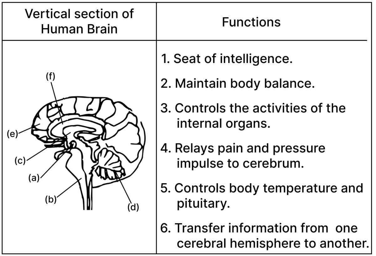
Given below is a vertical section of the human brain. Match the structures marked (a) to (e) with their correct functions.

Example: (f) – 6. Transfer information from one cerebral hemisphere to another.

Given below is a vertical section of the human brain. Match the structures marked (a) to (e) with their correct functions. ICSE Biology 2024 Specimen Paper Solved.
Show SolutionHide Solution↓

(a) → (5)

(b) → (3)

(c) → (4)

(d) → (2)

(e) → (1)

Explanation

(a) is Hypothalamus its function is to (5) Control body temperature and pituitary.

(b) is Medulla oblongata its function is to (3) Control the activities of the internal organs.

(c) is Thalamus its function is to (4) Relay pain and pressure impulse to cerebrum.

(d) is Cerebellum its function is to (2) Maintain body balance.

(e) is Cerebrum its function is the (1) Seat of intelligence.

3Long Answer Questions

State whether the following actions are voluntary actions, simple actions or conditioned reflex:

(i) Blinking

(ii) Cleaning the table

(iii) Playing on the keyboard

(iv) Salivating when food is put in the mouth

Show SolutionHide Solution↓

(i) Simple reflex (unconditioned reflex)

(ii) Voluntary action

(iii) Conditioned reflex

(iv) Simple reflex (unconditioned reflex)

4Long Answer Questions

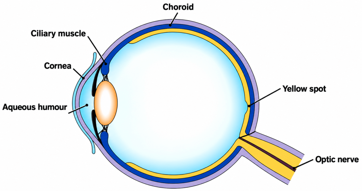
Draw a diagram of the human eye as seen in a vertical section and label the parts which suits the following functions/descriptions:

(i) The layer which prevents reflection of light.

(ii) The structure that alters the focal length of the lens.

(iii) The region of distinct vision.

(iv) The part which transmits the impulse to the brain.

(v) The outermost transparent layer in front of the eye lens.

(vi) The fluid present in the anterior part of the eye in front of the eye lens.

Show SolutionHide Solution↓

Below is the labelled diagram of the vertical section of the human eye:

Draw a diagram of the human eye as seen in a vertical section and label the parts which suits the following functions/descriptions:. The Nervous System & Sense Organs, ICSE Class 10 Biology by Goyal Brothers Solutions.

The parts suiting the given descriptions are:

(i) The layer which prevents reflection of light — Choroid

(ii) The structure that alters the focal length of the lens — Ciliary body (ciliary muscle)

(iii) The region of distinct vision — Yellow spot (fovea centralis)

(iv) The part which transmits the impulse to the brain — Optic nerve

(v) The outermost transparent layer in front of the eye lens — Cornea

(vi) The fluid present in the anterior part of the eye in front of the eye lens — Aqueous humour

5Long Answer Questions

Identify the odd one in each set and name the category to which the remaining three belong:

(i) Cyton, Photon, Axon, Dendron.

(ii) Haemoglobin, Glucagon, Iodopsin, Rhodopsin

(iii) Dendrites, Myelin Sheath, Axon, Spinal cord

(iv) Malleus, Incus, Cochlea, Stapes.

Show SolutionHide Solution↓

(i) Odd term: Photon Category: Parts of a neuron

(ii) Odd term: Glucagon Category: Pigments

(iii) Odd term: Spinal cord Category: Parts of a neuron

(iv) Odd term: Cochlea Category: Ear ossicles

6(i)Long Answer Questions

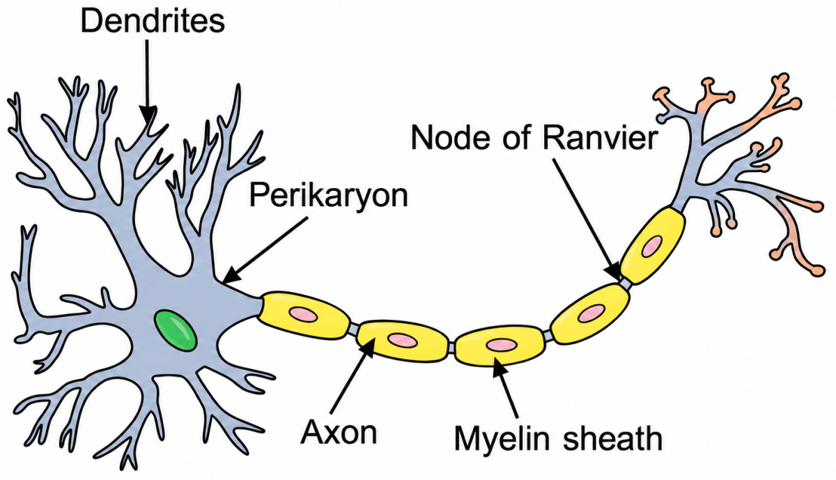
Draw a well-labelled diagram of a neuron showing the following parts : Perikaryon, Dendrites, Axon, Node of Ranvier and Myelin sheath.

Show SolutionHide Solution↓

Below is the well-labelled diagram of a neuron:

Draw a well-labelled diagram of a neuron showing the following parts: Perikaryon, Dendrites, Axon, Node of Ranvier and Myelin sheath. The Nervous System & Sense Organs, ICSE Class 10 Biology by Goyal Brothers Solutions.
6(ii)Long Answer Questions

State the function of sensory neuron and a motor neuron.

Show SolutionHide Solution↓

Sensory neuron — It transmits impulses from the receptors (sense organs) to the brain and spinal cord.
Motor neuron — It carries impulses (messages/instructions) from the brain and spinal cord to the effector organs such as muscles and glands.

6(iii)Long Answer Questions

What is a nerve made up of?

Show SolutionHide Solution↓

A nerve is made up of a bundle of nerve fibres (axons) of separate neurons, enclosed in a tubular sheath.

6(iv)Long Answer Questions

What is the function of ear ossicles?

Show SolutionHide Solution↓

The three ear ossicles (malleus, incus and stapes) transmit the vibrations from the ear drum to the oval window of the inner ear and amplify (magnify) them due to their lever-like action.

6(v)Long Answer Questions

How are the cytons and axons placed in the brain and the spinal cord?

Show SolutionHide Solution↓

In the brain (cerebrum), the cytons (cell bodies) lie in the outer region (cortex) forming the grey matter, while the axons lie in the inner region forming the white matter. In the spinal cord, the arrangement is reversed — the cytons (grey matter) lie on the inner side and the axons (white matter) lie on the outer side.

6(vi)Long Answer Questions

Which part of the human ear gives 'Dynamic balance' and 'Static balance' to the body?

Show SolutionHide Solution↓

Dynamic balance — Semicircular canals. Static balance — Vestibule (utriculus and sacculus).

6(vii)Long Answer Questions

Explain how the human eye adapts itself to bright light and dim light.

Show SolutionHide Solution↓

In dim light (dark adaptation), the pupil is dilated by the radial muscles of the iris to allow more light to enter, and the pigment rhodopsin is regenerated in the rods so that we can see in the dim light.

In bright light (light adaptation), the pupil is constricted by the circular muscles of the iris to reduce the amount of light entering, and the pigment iodopsin becomes active in the cones for clear vision in bright light. This adjustment of the eye to changing light intensities is called adaptation.

7Long Answer Questions

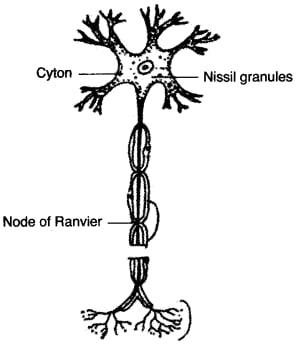
Draw a well labelled diagram of a 'Neuron' and name the following parts:
(a) Node of Ranvier
(b) Nissil granules
(c) Cyton

Show SolutionHide Solution↓

Well labelled diagram of a Neuron with Node of Ranvier, Nissil granules and Cyton named is shown below:

Draw a well labelled diagram of a Neuron and name the following parts - Node of Ranvier, Nissil granules, Cyton. ICSE 2012 Biology Solved Question Paper.
8(i)Long Answer Questions

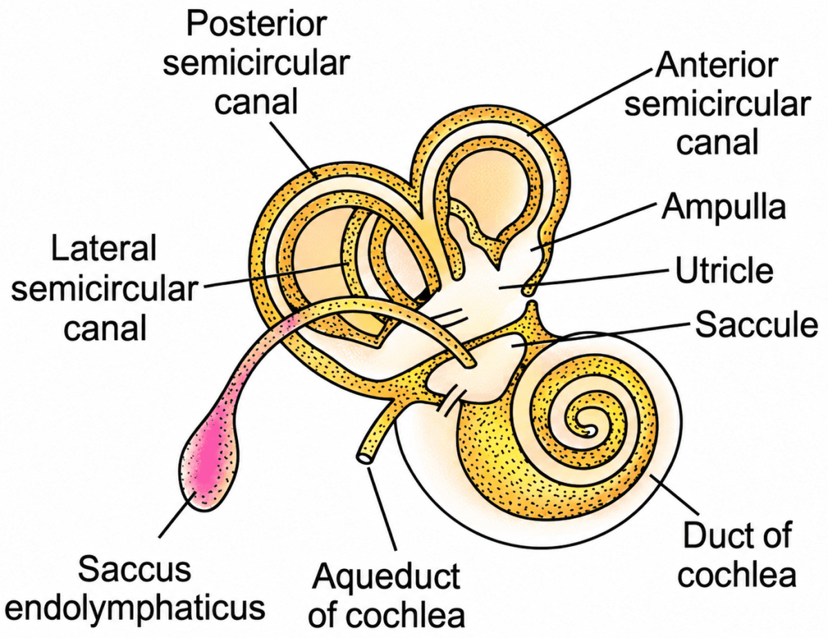
Draw a well-labelled diagram of the membranous labyrinth found in the inner ear.

Show SolutionHide Solution↓

Below is the well-labelled diagram of the membranous labyrinth found in the inner ear:

Draw a well-labelled diagram of the membranous labyrinth found in the inner ear. The Nervous System & Sense Organs, ICSE Class 10 Biology by Goyal Brothers Solutions.
8(ii)Long Answer Questions

Based on the diagram drawn above in (i) give a suitable term for each of the following descriptions:

(a) The sensory cells that helps in hearing.

(b) The part that is responsible for static balance of the body.

(c) The fluid present in the middle chamber of cochlea.

(d) The structure that maintains dynamic equilibrium of the body.

Show SolutionHide Solution↓

(a) Organ of Corti (sensory hair cells)

(b) Vestibule (utriculus and sacculus)

(c) Endolymph

(d) Semicircular canals

9Long Answer Questions

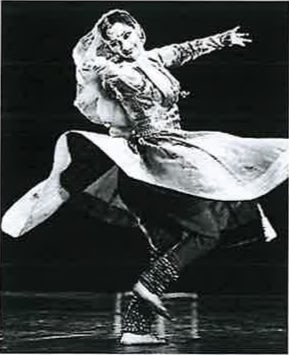
Kathak is an Indian classical dance which involves a lot of spinning by the dancers. The learners of this dance experience dizziness in the beginning while spinning.

Kathak is an Indian classical dance which involves a lot of spinning by the dancers. The learners of this dance experience dizziness in the beginning while spinning. ICSE 2025 Biology Solved Question Paper.

(a) Name the part of the membranous labyrinth responsible for this dizziness.

(b) Give a suitable reason for your answer.

(c) Mention the nerve that carries the impulse for dizziness to the brain.

Show SolutionHide Solution↓

(a) Semicircular canal

(b) The fluid in the semi-circular canals continue to spin for a short time even after the dancer stops. This results in dizziness.

(c) Auditory nerve

10Long Answer Questions

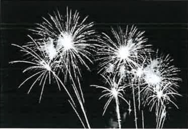
Chris was watching the display of fireworks in the sky.

Chris was watching the display of fireworks in the sky. ICSE 2025 Biology Solved Specimen Paper.

(a) Trace the path of the light rays using the following terms:

Fovea, Lens, Conjunctiva, Pupil, Cornea

(b) Name the nerve that carries the impulse for vision to the brain.

Show SolutionHide Solution↓

(a) Conjunctiva → Cornea → Pupil → Lens → Fovea

(b) Optic Nerve.

11(i)Long Answer Questions

Given below is a set of terms.

Medullary Sheath, Myelin Sheath, Dendron, Meninges, Nissl’s granules

(a) Choose the odd term from the above list.

(b) Mention the category to which the other terms belong.

Show SolutionHide Solution↓

(a) The odd term is Meninges.

(b) The other terms belong to parts or structures of a nerve cell (neuron).

11(ii)Long Answer Questions

Name the structure responsible for each of the following:

(a) conversion of mechanical sound waves to sound vibrations in the human ear.

(b) maintaining equal air pressure on either side of the tympanum.

Show SolutionHide Solution↓

(a) Ear drum (tympanic membrane)

(b) Eustachian tube

1Structured Questions

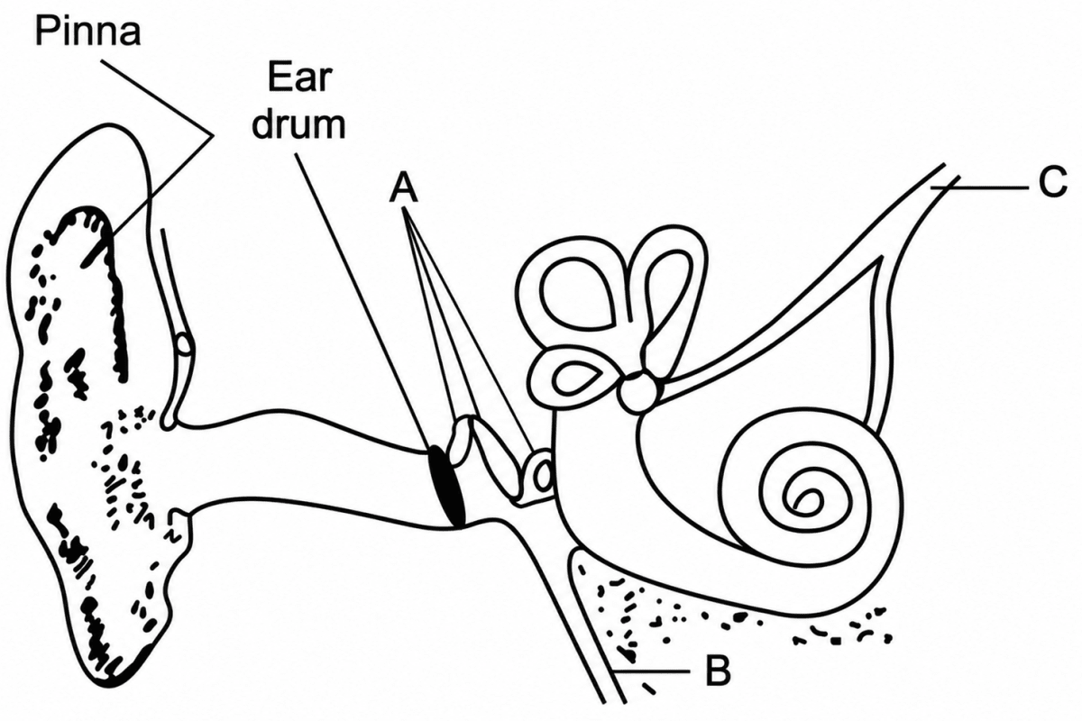
Given below is the diagram of the human ear. Study the same and answer the questions that follow:

Given below is the diagram of the human ear. Study the same and answer the questions that follow:. The Nervous System & Sense Organs, ICSE Class 10 Biology by Goyal Brothers Solutions.

(i) Give the biological term for the part labelled 'A' and state its function.

(ii) Name the part labelled 'B' and state its function.

(iii) Name the part labelled 'C' and state its function.

(iv) Give the function of ear wax.

Show SolutionHide Solution↓

(i) A → Ear ossicles (malleus, incus and stapes). Function — They transmit the vibrations from the ear drum to the oval window and amplify them due to their lever-like action.

(ii) B → Eustachian tube. Function — It connects the middle ear with the pharynx and equalises the air pressure on either side of the ear drum, allowing it to vibrate freely.

(iii) C → Semicircular canals. Function — They help in maintaining the dynamic equilibrium (balance during movement) of the body.

(iv) Ear wax, secreted by wax-secreting glands in the external auditory meatus, prevents the entry of dust and foreign particles into the ear.

2Structured Questions

Given below is a diagram depicting a defect of the human eye, study the same and then answer the questions that follow:

Given below is a diagram depicting a defect of the human eye, study the same and then answer the questions that follow. Name the defect shown in the diagram. What are the two possible reasons that cause this defect? Name the type of lens used to correct this defect. With the help of a diagram show how the defect shown above is rectified using a suitable lens. ICSE 2016 Biology Solved Question Paper.

(i) Name the defect shown in the diagram.

(ii) What are the two possible reasons that cause this defect?

(iii) What kind of spectacle lens is used to correct this defect?

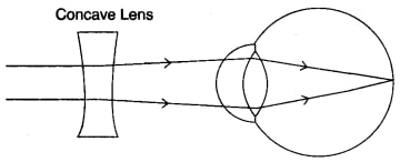
(iv) With the help of a diagram show how the defect shown above is rectified using a suitable spectacle.

Show SolutionHide Solution↓

(i) Myopia / short sightedness.

(ii) Two possible reasons that cause Myopia are:

  1. Eye ball is lengthened from front to back.
  2. Lens is too curved / rounded

(iii) Concave lens

(iv) Below diagram shows how a concave lens can be used to rectify Myopia:

With the help of a diagram show how the defect shown above is rectified using a suitable lens. ICSE 2016 Biology Solved Question Paper.
3Structured Questions

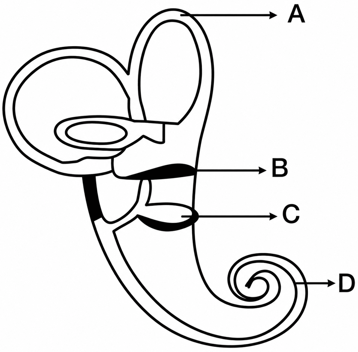
The diagram given below represents the structure found in the inner ear.

The diagram given below represents the structure found in the inner ear. Study the same and then answer the questions that follow:. The Nervous System & Sense Organs, ICSE Class 10 Biology by Goyal Brothers Solutions.

Study the same and then answer the questions that follow:

(i) Name the parts labelled A, B, C and D.

(ii) Name the part of the ear responsible for transmitting impulses to the brain.

(iii) Name the part labelled above which is responsible for:

(a) Static equilibrium

(b) Dynamic equilibrium

(c) Hearing.

(iv) Name the audio receptor cells which pick up vibrations.

(v) Name the fluid present in the inner ear.

Show SolutionHide Solution↓

(i) The labelled parts are:

A → Semicircular canals

B → Utriculus

C → Sacculus

D → Cochlea

(ii) Auditory nerve

(iii) The parts responsible are: (a) Static equilibrium — Utriculus and sacculus (vestibule) (b) Dynamic equilibrium — Semicircular canals (c) Hearing — Cochlea

(iv) Sensory hair cells of the organ of Corti

(v) Endolymph and perilymph

4Structured Questions

Viplav is a photographer. He finds that his vision gets blurred. This happens when he goes from taking pictures of faraway objects to reading text on his camera screen. He recalls learning about certain defects of the eyes during his school days.

Viplav is a photographer. He finds that his vision gets blurred. This happens when he goes from taking pictures of faraway objects to reading text on his camera screen. He recalls learning about certain defects of the eyes during his school days. ICSE 2025 Biology Solved Question Paper.

(a) Name the defect of the eyes which Viplav is suffering from.

(b) Mention the type of lens that can be used to correct the above defect.

Show SolutionHide Solution↓

(a) Hyperopia.

(b) Convex lens.

5Structured Questions

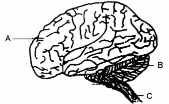
The diagram given below is an external view of the human brain. Study the same and answer the questions that follow:

The diagram given below is an external view of the human brain. Study the same and answer the questions. Name the parts labelled A, B and C in the diagram. State the main functions of the parts labelled A and B. What are the structural and functional units of the brain? How are the parts of these units arranged in A and C? Mention the collective term for the membranes covering the brain. What is the function of Cerebrospinal fluid? ICSE 2017 Biology Solved Question Paper.

(i) Name the parts labelled A, B and C in the diagram.

(ii) State the main functions of the parts labelled A and B.

(iii) What are the structural and functional units of the brain? How are the parts of these units arranged in A and C?

(iv) Mention the collective term for the membranes covering the brain.

(v) What is the function of Cerebrospinal fluid?

Show SolutionHide Solution↓

(i) The parts labelled A, B and C are:

  • A → Cerebrum
  • B → Cerebellum
  • C → Spinal Cord

(ii) The functions are mentioned below:

  • Cerebrum (Part A) — It is the seat of intelligence, consciousness and will power. It controls all voluntary activities.
  • Cerebellum (Part B) — It coordinates muscular activities and maintains balance of the body.

(iii) Neurons / nerve cells are the structural and functional units of the brain.
In part A (Cerebrum), Outer grey matter has cytons and inner white matter has axons.
In part C (Spinal Cord), Outer white matter has axons and inner grey matter has cytons.

(iv) Meninges

(v) Cerebrospinal fluid(CSF) acts like a cushion to protect the brain / spinal cord from injuries and shocks.

6Structured Questions

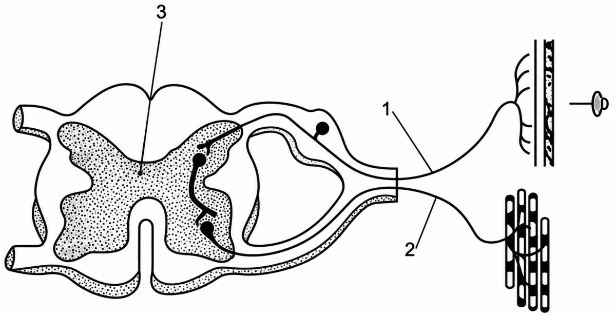
The diagram given below shows the internal structure of a spinal cord depicting a phenomenon. Study the diagram and answer the questions:

The diagram given below shows the internal structure of a spinal cord depicting a phenomenon. Study the diagram and answer the questions:. The Nervous System & Sense Organs, ICSE Class 10 Biology by Goyal Brothers Solutions.

(i) Define the phenomenon that is depicted in the diagram.

(ii) Give the technical term for the gap between two neurons.

(iii) Define the parts numbered 1, 2 and 3.

(iv) Discuss the arrangement of white & gray matter in brain and spinal cord.
(v) Mention two ways by which the spinal cord is protected in our body.

Show SolutionHide Solution↓

(i) The phenomenon depicted is reflex action. It is a rapid, automatic, involuntary response to a stimulus, for example, immediate withdrawal of the hand on touching a hot object.

(ii) Synapse (synaptic cleft)

(iii) The parts numbered 1, 2 and 3 are:

1 → Sensory (afferent) neuron — the neuron that carries impulses from the receptor to the spinal cord.

2 → Motor (efferent) neuron — the neuron that carries impulses from the spinal cord to the effector (muscle or gland).

3 → Grey matter — the inner, H-shaped region of the spinal cord containing the cytons (cell bodies) and connecting neurons.

(iv) In the brain (cerebrum), the grey matter (cytons) lies on the outer side and the white matter (axons) lies on the inner side. In the spinal cord, the arrangement is reversed — the white matter surrounds the grey matter, i.e. the grey matter (cytons) lies on the inner side and the white matter (axons) lies on the outer side.

(v) The spinal cord is protected by:

  1. The bony vertebral column (backbone) in which it lies.
  2. The three meninges (dura mater, arachnoid and pia mater) and the cerebrospinal fluid, which act as a shock-absorbing cushion.

Press ↓ to reveal a solution and move to the next question. Back to Goyal New Approach Biology.

Free sample bank